de-Leon, B. - T. S. ; Oren, R. ; Spira, M. E. ; Korbakov, N. ; Yitzchaik, S. ; Saar, A. Porous Silicon Substrates for Neurons Culturing and Bio-Photonic Sensing.
Physica Status Solidi A 2005,
202, 1456-1461.
ArticleAbstractIn this work we report on culturing of Aplysia neurons and vertebrate cells to porous silicon substrates and on the first steps toward characterizing porous silicon as a biosensor of neural activity. Neurons cultured on porous silicon substrates survived for at least one week showing normal passive membrane properties and generation of action potentials. We have investigated several mechanisms that take advantage of the optical properties of porous silicon for transducing both electrical and chemical neuronal activities into photonic signals. For example, the photoluminescence response to voltage and the reflectivity response to chemical changes were investigated.
Ofir, Y. ; Schwartsglass, O. ; Shappir, J. ; Yitzchaik, S. Organic Field Effect Transistors Based on Multilayer Films via Molecular Epitaxy.
Organic Thin-Film Electronics 2005,
871.
ArticleAbstractA Self-Assembly oriented technique from the vapor-phase, Molecular Layer Epitaxy (MLE), was utilized for the buildup of organic multilayers as the active channel in organic field effect transistors (OFET). Carrier gas-assisted chemical vapor deposition (C
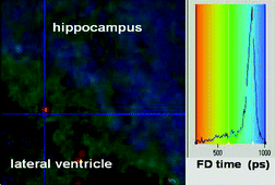
Vaganova, E. ; Yitzchaik, S. ; Sigalov, M. ; Khodorovsky, V. ; Borst, J. W. ; Visser, A. ; Ovadia, H. Time-Resolved Emission upon Two-Photon Excitation of bis-N-Carbazolyl-distyrylbenzene: Mapping of Water Distribution in the Mouse Brain.
New Journal of Chemistry 2005,
29, 1044-1048.
ArticleAbstractWe present a method of mapping the water molecule distribution in mouse brain tissues using injected bis-N-carbazolyl-distyrylbenzene and the FLIM technique. The fluorescence lifetime of this two-photon absorbing chromophore diminishes when the amount of water in the surrounding area increases. The fluorescence lifetime of the injected in vivo chromophorestrongly depends on the content of water in different areas. Thus, lifetimes of 900 ± 50 ps in the hippocampus (extracellular fluid), 520 ± 50 ps in the lateral ventricle (choroid plexus, cerebrospinal fluid), and 400–150 ps in blood vessels were observed. Moreover, the fluorescence lifetime distribution undergoes drastic changes when mice are deprived of water. Statistical analysis of the investigated samples showed that upon water deprivation watercontent decreased at the border of the hippocampus/lateral ventricle areas and increased in blood vessels.